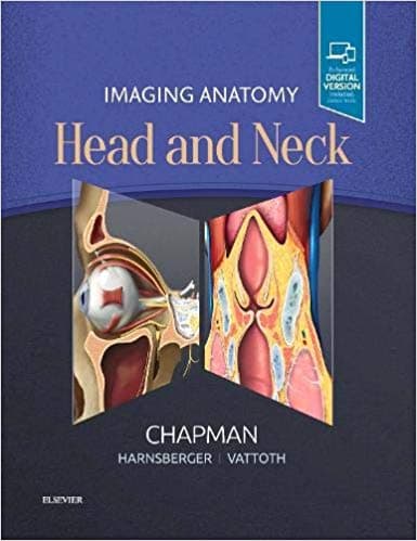
<p>Highly specialized structures, microanatomy of individual components, and overall structural density make the head and neck one of the most challenging areas in radiology. <font color="#333333" face="Arial, sans-serif"><span style="font-size: 14px;">Imaging Anatomy: Head and Neck</span></font> provides radiologists, residents, and fellows with <font color="#333333" face="Arial, sans-serif"><span style="font-size: 14px;">a truly comprehensive, superbly illustrated anatomy reference </span></font>that is designed to improve interpretive skills in this complex area. A wealth of <font color="#333333" face="Arial, sans-serif"><span style="font-size: 14px;">high-quality, cross-sectional images, corresponding medical illustrations, and concise, descriptive text</span></font> offer a unique opportunity to master the fundamentals of normal anatomy and accurately and efficiently recognize pathologic conditions.</p><ul style="margin-right: 0px; margin-left: 20px; padding: 0px; margin-bottom: 0px !important;"><li style="margin: 0px 0px 0px 20px; overflow-wrap: break-word;">Contains more than 1,400 high-resolution, cross-sectional head and neck images combined with over 200 vibrant medical illustrations, designed to provide the busy radiologist rapid answers to imaging anatomy questions</li><li style="margin: 0px 0px 0px 20px; overflow-wrap: break-word;">Reflects new understandings of anatomy due to ongoing anatomic research as well as new, advanced imaging techniques</li><li style="margin: 0px 0px 0px 20px; overflow-wrap: break-word;">Features 3 Tesla MR imaging sequences and state-of-the-art multidetector CT normal anatomy sequences throughout the book, providing detailed views of anatomic structures that complement highly accurate and detailed medical illustrations</li><li style="margin: 0px 0px 0px 20px; overflow-wrap: break-word;">Includes imaging series of successive slices in each standard plane of imaging (coronal, sagittal, and axial)</li><li style="margin: 0px 0px 0px 20px; overflow-wrap: break-word;">Depicts anatomic variations and pathological processes to help you quickly recognize the appearance and relevance of altered morphology</li><li style="margin: 0px 0px 0px 20px; overflow-wrap: break-word;">Includes CT and MR images of pathologic conditions, when appropriate, as they directly enhance current understanding of normal anatomy</li><li style="margin: 0px 0px 0px 20px; overflow-wrap: break-word;">Contains a separate section on normal ultrasound anatomy of the head and neck</li><li style="margin: 0px 0px 0px 20px; overflow-wrap: break-word;"><font color="#333333" face="Arial, sans-serif"><span style="font-size: 14px;">Expert Consult™ eBook version included with purchase</span></font>, which allows you to search all of the text, figures, and references from the book on a variety of devices</li></ul>