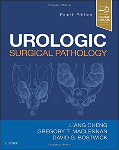
<p style="margin-bottom: 14px; padding: 0px; color: rgb(51, 51, 51); font-family: Arial, sans-serif; font-size: 14px;">Offering comprehensive coverage of this fast-changing field for more than 20 years, <i style="">Urologic Surgical Pathology</i> is an expert guide to all common and rare entities in the genitourinary system. The 4th Edition keeps you fully up to date with discussions of newly recognized tumors and terminologies, the latest classification schemes, current grading approaches, molecular alterations, and commonly used ancillary diagnostic techniques. With its clinical focus on day-to-day urological pathology sign-outand an emphasis on clinicopathologic and radiographic-pathologic correlations, this thoroughly revised uropathology reference is an excellent resource for diagnostic decision making.<i></i></p><ul style="margin-right: 0px; margin-left: 20px; padding: 0px; color: rgb(51, 51, 51); font-family: Arial, sans-serif; font-size: 14px; margin-bottom: 0px !important;"><li style="margin: 0px 0px 0px 20px; overflow-wrap: break-word;">Includes expanded coverage of differential diagnosis for all tumor types encountered in urological surgical pathology practice.</li><p style="margin-bottom: 14px; padding: 0px;"></p><li style="margin: 0px 0px 0px 20px; overflow-wrap: break-word;">Incorporates the latest TNM staging and WHO classification systems, as well as new diagnostic biomarkers and their utility in differential diagnosis, newly described variants and new histologic entities.</li><p style="margin-bottom: 14px; padding: 0px;"></p><li style="margin: 0px 0px 0px 20px; overflow-wrap: break-word;">Discusses advances in molecular diagnostic testing, its capabilities, and its limitations, including targeted therapy/personalized medicine.</li><p style="margin-bottom: 14px; padding: 0px;"></p><li style="margin: 0px 0px 0px 20px; overflow-wrap: break-word;">Covers new developments in immunohistochemistry and the latest diagnostic tumor markers.</li><p style="margin-bottom: 14px; padding: 0px;"></p><li style="margin: 0px 0px 0px 20px; overflow-wrap: break-word;">Features more than 1,600 high-quality images – all in color – including gross pictures, histopathologic and cytopathologic images, special stains, other ancillaries, drawings, and illustrations.</li><p style="margin-bottom: 14px; padding: 0px;"></p><li style="margin: 0px 0px 0px 20px; overflow-wrap: break-word;">Helps you find information quickly with a consistent chapter format; an abundance of tables, diagrams and flowcharts; boxed lists of types and causes of diseases; differential diagnosis; characteristic features of diseases; complications; classifications; and staging.</li><p style="margin-bottom: 0px; padding: 0px;"></p><li style="margin: 0px 0px 0px 20px; overflow-wrap: break-word;">Enhanced eBook version included with purchase. Your enhanced eBook allows you to access all of the text, figures, and references from the book on a variety of devices.</li></ul>